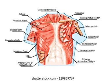
Home » Without Label » Anatomy Of Chest - Artstation Male Chest Anatomy Practice Tomas Sosto / It provides protection to vital organs (eg, heart and major vessels, lungs, liver) and provides stability for movement.
Anatomy Of Chest - Artstation Male Chest Anatomy Practice Tomas Sosto / It provides protection to vital organs (eg, heart and major vessels, lungs, liver) and provides stability for movement.
Anatomy Of Chest - Artstation Male Chest Anatomy Practice Tomas Sosto / It provides protection to vital organs (eg, heart and major vessels, lungs, liver) and provides stability for movement.. Sternocleidomastoid muscle clavicle and ribs anatomy muscle anatomy chest sternocleidomastoid ribs anatomy chest muscles anatomy thorax rib muscles chest muscles chest anatomy illustration. It provides protection to vital organs (eg, heart and major vessels, lungs, liver) and provides stability for movement. Several muscles that move the arms, head, and neck have their origins on the sternum. Table 1.1 lists the major anatomic structures within the thorax that are discussed. 31 anatomy of the female breast syllabus p.
The anatomic illustrations are presented as… Related posts of anatomy of the chest abdominal blood supply. About the 6th week, the somites differentiate into the sclerotomes and the dermatomyotomes. Structures to identify • heart • lungs • mediastinum • pleural space • chest wall • …everything else! In insects, crustaceans, and the extinct trilobites, the thorax is one of the three main divisions of the creature's body, each of which is in turn composed of multiple segments.
Human Chest Anatomy Photograph By Leonello Calvetti Science Photo Library from images.fineartamerica.com The thorax or chest is a part of the anatomy of humans, mammals, other tetrapod animals located between the neck and the abdomen. A good radiologist knows the anatomy because knowing where structures normally live and recognizing the location of an abnormality helps to make or narrow the differential diagnosis. Plus, how to target each to make them bigger and stronger. The myotomes elongate and invade the mesoderm of the wall of the embryonic thoracic and abdominal cavities. It is important to remember the position and orientation of the heart when placing a stethoscope on the chest of a patient and listening for heart sounds, and also when looking at images taken from a midsagittal perspective. The first step in understanding thorax anatomy is to find out its boundaries. Anatomically, the heart is located in the anterior thoracic cavity; Learn about each of these muscles, their locations, functional anatomy and exercises for them.
2 skin of the anterior chest wall syllabus p.
The clavicular head, the sternal head, and the abdominal head. This chapter is an abbreviated review of thoracic anatomy as seen on chest radiographs and computed tomography (ct) of the chest. This atlas is a comprehensive and affordable learning tool for medical students and residents and especially for radiologists and pneumologists. It is enclosed by the ribs, the vertebral column, and the sternum, or breastbone, and is separated from the abdominal cavity (the body's largest hollow space) by a muscular and membranous partition, the diaphragm. The right side of the heart is deflected anteriorly, and the left side is deflected posteriorly. About the 6th week, the somites differentiate into the sclerotomes and the dermatomyotomes. The first step in understanding thorax anatomy is to find out its boundaries. The trapezius originates from the skull and spine of the upper back and neck. Browse 6,436 chest anatomy stock photos and images available, or search for human anatomy to find more great stock photos and pictures. The chest is the area of origin for many of the body's systems as it houses organs such as the heart, esophagus, trachea, lungs, and thoracic diaphragm. It is important to remember the position and orientation of the heart when placing a stethoscope on the chest of a patient and listening for heart sounds, and also when looking at images taken from a midsagittal perspective. Chest a man's chest — like the rest of his body — is covered with skin that has two layers. It provides access to ct images in the axial plane, allowing the user to learn and review the lung anatomy interactively.
Basic thoracic anatomy and physiology an understanding of thoracic imaging requires knowledge of the anatomy being imaged, as described in this chapter, as well as the imaging techniques applied to the thorax, covered in chapter 2. Computed tomography (ct) of the chest can detect pathology that may not show up on a conventional chest radiograph(1). This chapter is an abbreviated review of thoracic anatomy as seen on chest radiographs and computed tomography (ct) of the chest. Learn about each of these muscles, their locations, functional anatomy and exercises for them. The sternum, commonly known as the breastbone, is a long, narrow flat bone that serves as the keystone of the rib cage and stabilizes the thoracic skeleton.
Human Chest Anatomy Images Stock Photos Vectors Shutterstock from image.shutterstock.com This atlas is a comprehensive and affordable learning tool for medical students and residents and especially for radiologists and pneumologists. The pectoralis major and the pectoralis minor, known collectively as your pecs. Muscles of the chest and their functions you have two mighty muscles on both sides of your chest: 2 skin of the anterior chest wall syllabus p. Table 1.1 lists the major anatomic structures within the thorax that are discussed. Use the mouse scroll wheel to move the images up and down alternatively use the tiny arrows (>>) on both side of the image to move the images.>>) on both side of the image to move the images. The clavicular head, the sternal head, and the abdominal head. The trapezius originates from the skull and spine of the upper back and neck.
The anatomic illustrations are presented as…
Bones of the chest and upper back understanding chest wall anatomy is paramount to any surgical procedure regarding the chest and is vital to any reco. The circulatory system does most of its work. The anatomy of the sternum. This chapter is an abbreviated review of thoracic anatomy as seen on chest radiographs and computed tomography (ct) of the chest. See chest anatomy stock video clips. These heads are important to know because they can be specifically trained through particular movements. Muscles of the chest and their functions you have two mighty muscles on both sides of your chest: The right side of the heart is deflected anteriorly, and the left side is deflected posteriorly. These myotomes divide into the epimere and the hypomere. Anatomy of right side chest pain. 4 innervation of the breast blood supply of the breast syllabus p. It provides access to ct images in the axial plane, allowing the user to learn and review the lung anatomy interactively. The trapezius originates from the skull and spine of the upper back and neck.
The circulatory system does most of its work. 30 lines of the thoracic wall syllabus p. Sternocleidomastoid muscle clavicle and ribs anatomy muscle anatomy chest sternocleidomastoid ribs anatomy chest muscles anatomy thorax rib muscles chest muscles chest anatomy illustration. Use the mouse scroll wheel to move the images up and down alternatively use the tiny arrows (>>) on both side of the image to move the images.>>) on both side of the image to move the images. 4 innervation of the breast blood supply of the breast syllabus p.
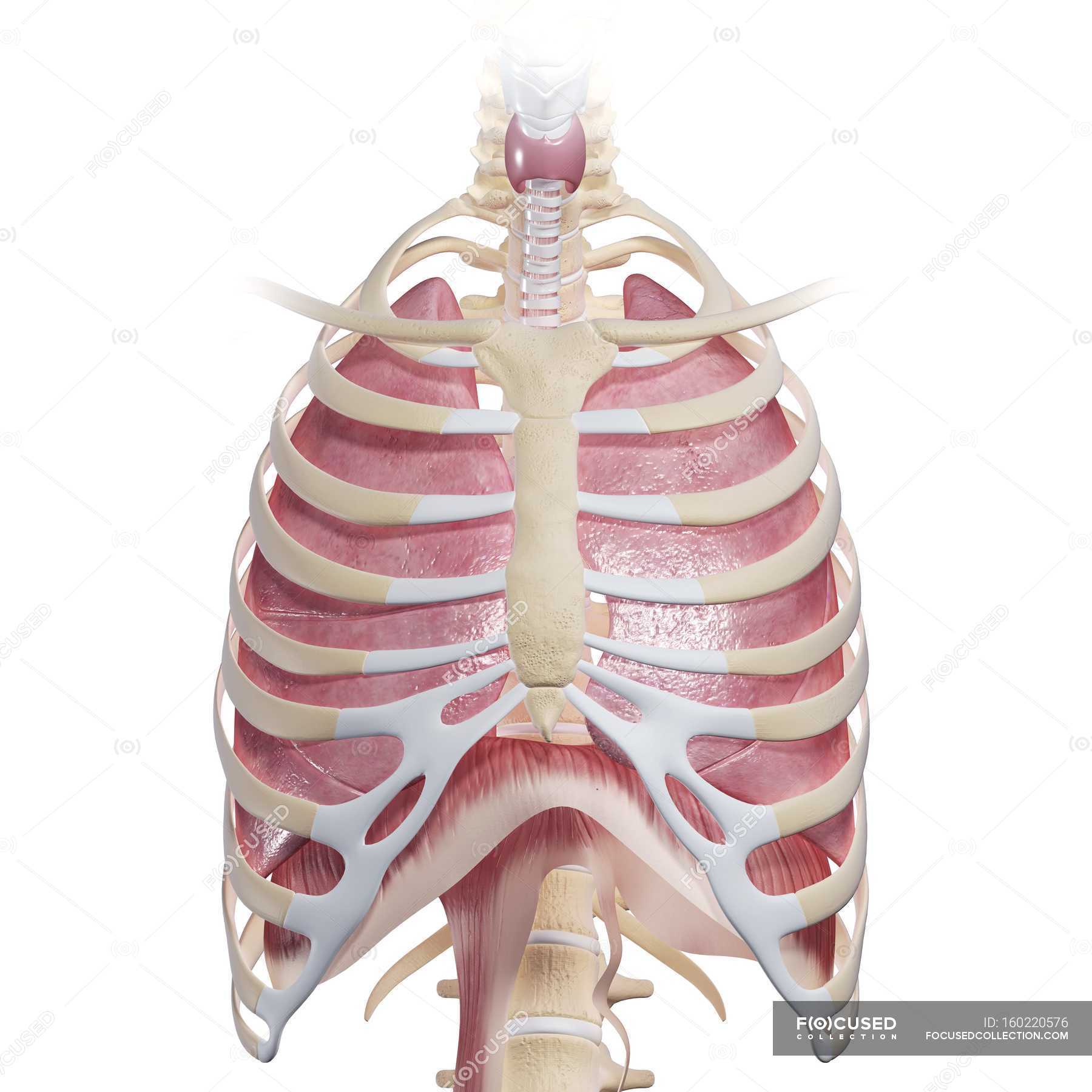
Human Chest Anatomy Larynx Physiology Stock Photo 160220576 from st.focusedcollection.com The chest is the area of origin for many of the body's systems as it houses organs such as the heart, esophagus, trachea, lungs, and thoracic diaphragm. Learn about each of these muscles, their locations, functional anatomy and exercises for them. It provides access to ct images in the axial plane, allowing the user to learn and review the lung anatomy interactively. The chest or thorax region of the upper body has a number of important organs that reside within it that may present with chest pain if they become compromised in. The anatomy of the sternum. About the 6th week, the somites differentiate into the sclerotomes and the dermatomyotomes. System respiratory respiratory organs of human body digestive and respiratory system medical chest internal structure of human body medicine body lungs biology intestines stomach anatomy torso human internal. The clavicular head, the sternal head, and the abdominal head.
Get the full built by science program:
The thorax or chest is a part of the anatomy of humans, mammals, other tetrapod animals located between the neck and the abdomen. The pectoralis major and the pectoralis minor, known collectively as your pecs. Related posts of anatomy of the chest abdominal blood supply. See chest anatomy stock video clips. The sternum, commonly known as the breastbone, is a long, narrow flat bone that serves as the keystone of the rib cage and stabilizes the thoracic skeleton. The chest or thorax is the region between the neck and diaphragm that encloses organs, such as the heart, lungs, esophagus, trachea, and thoracic diaphragm. This chapter is an abbreviated review of thoracic anatomy as seen on chest radiographs and computed tomography (ct) of the chest. The anatomy of the sternum. The clavicular head, the sternal head, and the abdominal head. It also protects several vital organs of the chest, such as the heart, aorta, vena cava, and. It provides protection to vital organs (eg, heart and major vessels, lungs, liver) and provides stability for movement. The first step in understanding thorax anatomy is to find out its boundaries. The trapezius originates from the skull and spine of the upper back and neck.